|
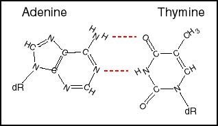
The base pairs In the double stranded DNA found in cells, bases occur as two pairs: guanine with cytosine, and adenine with thymine. This pairing is due to specific hydrogen bonding between the bases (the red dashed lines), and is the basis of the ability of DNA to replicate. Each pair consists of a two ring base (a purine) and a base with only one ring (a pyrimidine). Thus the size of each pair, and particularly the spacing between the two deoxyribose sugars is almost identical. The bases are linked to deoxyribose (dR in the figures) by a nitrogen-carbon glycosidic bond. |
|||||||||
|
The bases are flat As you trace around the rings, note that each carbon has one double bound, so the three atoms linked to the carbon are in a plane, and the entire ring structure is planar. The planar nature of the bases will be seen later to be central to the structure of the DNA helix. The alternating single and double bonds in the rings results in absorption in the u.v. region of the spectrum. |
|||||||||
 |
|||||||||
|
A:T is weaker than G:C The adenine-thymine base pair is formed with two hydrogen bonds, and is thus slightly weaker than the guanine-cytosine pair. DNA molecules containing a high percent of A:T pairs are typically less stable to high temperature than DNA with more G:C pairs. |
 |
||||||||
 |
Space filling model of C:G To the left we see the C:G nucleotide (base + sugar + phosphate) pair on our home page. This is a more realistic picture of the atomic electron distribution and thus the volume other atoms are excluded from. On the other hand, some atoms are obscured by others. However, now we are experts on nucleotide structure and can find our way around the molecule. |
||||||||
| Starting on the outsides, the large yellow balls with red bumps are the two phosphate atoms and their oxygens. Working toward the middle, the two riboses are not too impressive, as the small white hydrogen and light gray carbons tend to fuse together. We can see one of the red ribose oxygens on the left, and two on the right. The first purple triangle is the nitrogen of the glycosidic bond. The six atom cytosine ring on the left is obvious, as is the six:five ring of the guanine on the right (I oriented the molecule so the bases would be flat). Finally, you can see only two small black gaps between the three hydrogen bonds that make the pair, with the red oxygen of guanine at the top, and the cytosine oxygen on the bottom. | |||||||||