Bacteria

Most bacteria (a few species are photosynthetic) can't get energy from the sun or make food from CO2 and water. Bacteria are scavengers, microbial vultures. However they can't actually eat carcasses, the food must be first be degraded to small organic molecules by various enzymes, e.g. proteases, nucleases, lipases.
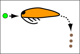
To see the role of bacteria in one section of our food pyramide look at the cartoon of a copepod (side view) on the left . The copepod is eating food, here a green dinoflagellate that has eaten bacteria and diatoms. As the food works its way down the gut of the copepod it is digested into small molecules by enzymes secreated by cells of the copepod. Only food that is degraded into small molecules can be adsorbed and used to make copepod tissue and oxidized to generate energy.

However, the gut of the copepod also contains a large population of bacteria that also secrete enzymes that help degrade the food the copepod has eaten. Of course the bacterial adsorb some of the nutriants they help generate. As the bacteria grow and multiply many end up, along with undigested food, in the stream of defecated material, the brown fecal pellets exiting the copepod on the right.

Where are the bacteria in this picture? I didn't draw the bacteria because they would be so small you wouldn't be able to see them (they are less than 1/100 th the size of the copepod). It is also redundant to draw them because they are everywhere in the picture. Bacteria are in the dinoflagelate, they are in the copepod's gut, they are in the fecal pellets, and they are also drifting around in the water that the other organisms are floating in. There would be different mixes of bacterial species in different locations, but in the Figure they would all look like spots. Bacteria are so ubiquitous that they can be considered part of the environment. However, it is only in the last decade that we have really begun to understand the magnitude of the bacterial population. It's not as easy to detect bacteria as you might imagine.

Finding bacteria

No problem, use a microscope. Not so fast. If you use a medium power objective so you can scan a reasonable area of a microscope slide the bacterium will appear as dark specks. You can't tell if they are really bacteria or if they are alive. In order to recognize a bacteria you need to see some detail inside the cell. However, to do this you need high power, say 100x, which also means you need oil between the objective and the slide (the yellow drop in the Figure). You also need fancy illumination (the red arrows) and stains that will reveal the inner parts of the cell. This is difficult enough if you have a concentrated bacterial suspension and want to characterize the cells. But it's really not a usefull technique to scan large volumes of fluid or complex suspensions of particles.
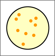
No problem, plate it out. This phrase refers to the most common way to test for bacteria. You smear the material to be tested on the top surface of a sterile nutrient gel which is contained in a Petri dish, or plate. The dish is then incubated for several days at the optimal temperature for bacterial growth. Any bacterium that can grow will produce a visable lump on the surface of the gel. This is because a typical bacterium can grow and divide every hour, so in one day one bacteria cell can produce 224 or 16,000,000 daughter cells. This quantity of material is visable to the eye as a colony (the dark orange spots on the Figure to the left). The colonies are clones: they all derive from a single cell.

The trouble is that you can only detect bacteria that are able to grow on the nutrient gel you provide. Colonies mean there are bacteria; no colonies do not mean there are no bacteria. Unfortunately, most bacteria floating around in the ocean do not grow on the typical nutrient gel used by bacteriologists.

The unlikely answer: DNA sequencing. Usually the nucleotide squence of the DNA of an organism is determined only after a great deal is known about the organism and it's considered so interesting that it's worth the time and money to find the sequence, e.g. the DNA sequence of humans. However, the cost of DNA sequencing is rapidly decreasing and the proceedure is now used in unexpected ways. Craig Venter and collegues sampled ocean water from the Sargasso Sea and just squenced every DNA molecule in the sample [Science v304p66(2004)]. The conclusion was that the sea water contained at least 100 times the number of bacteria that could be cultured by plating the water on nutrient media. Of course a great deal more information was obtained. One of the conclusions was that typical ocean water, that appears clear to the eye, contains about 107 bacteria, and 109 bacterial viruses per ml (more about viruses later).

Bacteria can only use small molecules as food: how are large molecules broken down to small ones?

Many bacteria secrete enzymes that degrade proteins, nucleic acids, and fats into smaller molecules that can be taken up by bacteria (bacteria have molecular pumps that can concentrate these small molecules even when present in the environment in very low concentrations).

In other cases the bacteria is in an environment where another organism provides the enzymes, e.g. the gut.

All cells contain enzymes that degrade proteins, fats, etc. In the life of a cell almost all molecules have a finite life time for optimal function. Enzymes in the cell then degrade the molecules and the smaller molecular fragments are reused to make new large molecules. This turnover process is under strict control, it's not just random chaos. However, when cells die or are injured, these enzymes are no longer under control, they become active and digest the cell. At the level of the whole organism, a level we can see without a microscope, dead plants and animals degrade themselves producing a soup of small molecles than can be used by bacteria.

All organisms live in association with bacteria, e.g. humans have bacteria on their skin, in their mouth and lungs, and throughout the digestive system. When we die our skin and endothelial cells quickly loose the ability to keep these bacteria out and we are consumed by them. That is the end and beginning of life.


How do bacteria die?

Most organisms have an intrinsic life span, but bacteria do not. They propagate by cell division; the two daughter cells appear to be identical and they divide in turn and so on and so on. We have mentioned that bacteria are eaten by dinoflagelates; as you might suspect many other organisms eat bacteria. Bacteria are also killed by many conditions in their environment, the UV in intense sunlight, and they die if deprived of food for long periods of time (but the resistance to starvation varies greatly from species to species).

All plants, animals, and bacteria are periodically infected by specific viruses. Viruses are thus a major mechanism for converting bacteria into small nutriant molecules that are consumed by other bacteria.

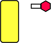
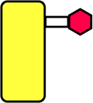
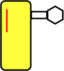
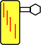
Viruses that attack bacteria are also called bacteriophages. A typical bacteriophage has a hexagnonal protein head containing its DNA genome (shown as red in the Figure to the right). However, a few bacteriophages an RNA genome.
The virus binds to the surface of its bacterial host using fibers at the end of its tail (the hollow rectangle attached to the red head).
The DNA (the red line) is injected into the bacterial cell while the empty protein shell is left on the surface. Hershey and Chase showed that you could shear off the empty shell at this stage of the infection and new bacteriophage would still be produced. This was one of the pivital experiments that demonstrated DNA was the genetic material.
Once inside the cell the virus DNA replicates using many of the bacterial enzymes. The viral DNA then is copied into RNA and then into new protein subunits which assemble around the DNA to make new virus particles.
The viral DNA also codes for enzymes that degrade the bacterial cell wall from the inside, and eventually the bacterial cell breaks open to release the new virus particles into the ocean (in our example).

<back to the food chain>