Figure A-5.
Four examples of colonial tunicates:
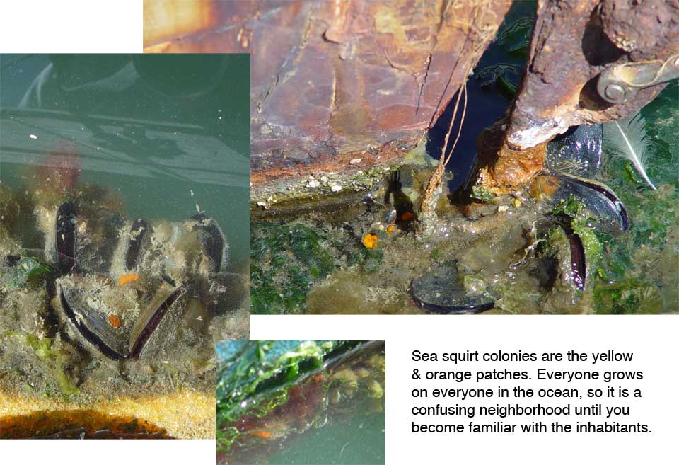
Bottom left panel, right side: Eight colonies of Botryllus sp. Each colony is a radial cluster of about 9 zooids, each with its own intake siphon, and a central shared exit siphon.
Top left and right, and bottom right panel: Colonies of several species of the genus Botrylloides. In this genus the zooids are typically aligned in rows, not circles. The placement of the exit siphon is irregular, and depends on the geometry of the colony. Arrangement of zooids is influenced by size and age of the colony as well as the geometry of the surface the colony is growing on. Thus there are colonies which are difficult to identify with one or the other genus.
There are other organisms here also. In the top right and bottom left panel you can see dark red (or purple) arms of the bryozoan Bugula neritina in the upper parts of the panel. In the lower left panel, bottom, you can see (if you know what to look for) the exit siphon of the almost completely transparent tunicate Diplosoma sp. There is just the faintest circular outline of its siphon, with its body revealed because it makes the background under it look out of focus (lower part of the panel).